
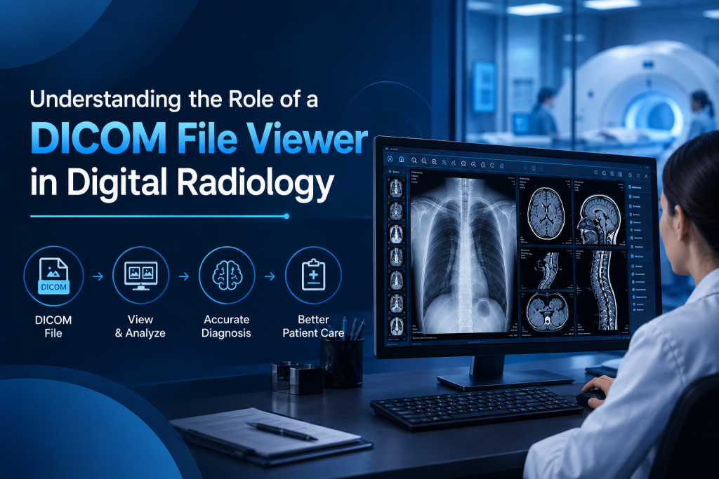
Digital radiology has transformed healthcare, enabling faster diagnoses, better collaboration, and efficient management of medical imaging data. Central to this transformation is the DICOM (Digital Imaging and Communications in Medicine) standard, which ensures that medical images from modalities like X-rays, CT scans, MRIs, and ultrasounds can be stored, transmitted, and interpreted consistently.
To fully leverage these images, radiologists rely on DICOM file viewers. These specialized software tools allow clinicians to view, manipulate, and analyze medical images, supporting accurate diagnoses and efficient workflow.
What Is a DICOM File Viewer?
A DICOM file viewer is a software application designed to read and display DICOM files. Unlike standard image formats like JPEG or PNG, DICOM files contain both the medical image and important metadata, such as patient information, acquisition parameters, and study details.
Key capabilities of a DICOM file viewer include:
- Displaying high-resolution images across multiple imaging modalities
- Adjusting contrast and brightness to highlight specific tissues
- Measuring and annotating areas of interest for diagnostic purposes
- Comparing current and prior studies to monitor disease progression
By offering these tools, a DICOM viewer ensures radiologists can accurately interpret complex medical images.
Supporting Multiple Imaging Modalities
Modern hospitals use a wide range of imaging techniques, and a robust DICOM file viewer supports multiple modalities, including:
- X-ray: Quick assessment of bones and chest conditions
- CT scans: Detailed cross-sectional views of internal organs
- MRI: High-resolution images of soft tissues, brain, and joints
- Ultrasound: Dynamic imaging for real-time assessment
Supporting multiple modalities within one viewer allows radiologists to handle all their studies efficiently without switching between different software applications.
Enhancing Diagnostic Accuracy
A DICOM file viewer enhances diagnostic accuracy through advanced visualization and analysis tools. Features such as zooming, panning, and windowing allow radiologists to examine fine details in high-resolution images.
Additional tools include:
- Multi-planar reconstruction (MPR): View anatomy from multiple planes for better understanding of complex structures
- Annotations and measurements: Provide precise documentation for lesions, fractures, or abnormalities
- Image fusion: Compare and overlay images from different modalities for comprehensive evaluation
These capabilities enable radiologists to make informed decisions, reducing the likelihood of errors and improving patient outcomes.
Improving Workflow Efficiency
Radiology departments often manage large volumes of imaging studies. A DICOM file viewer improves workflow efficiency by:
- Integrating with PACS and RIS: Automatically retrieves patient studies and updates reports
- Prioritizing cases: Worklists and tagging allow urgent studies to be addressed first
- Streamlining reporting: Measurements and annotations can be transferred directly to reports
By simplifying these tasks, radiologists spend more time analyzing images and less time managing technical and administrative work.
Enabling Remote and Teleradiology Reporting
Teleradiology has become a critical aspect of modern healthcare, allowing radiologists to read studies remotely. A DICOM file viewer supports remote reporting by:
- Providing secure access to cloud or server-based images
- Ensuring consistent image quality across devices
- Allowing collaboration with colleagues through image sharing and annotation
This capability is particularly valuable for rural hospitals, multi-location facilities, or after-hours reporting, ensuring timely diagnoses and continuous patient care.
Supporting Education and Training
DICOM viewers are also important tools for education and training in radiology. Trainees can learn from annotated cases, multi-planar images, and multimodality studies. Features like side-by-side comparison and image fusion provide practical experience in interpreting complex cases, preparing the next generation of radiologists for real-world challenges.
Integration with Advanced Technologies
Modern DICOM file viewers often integrate with advanced technologies such as AI-assisted analysis and automated detection tools. These features help radiologists:
- Highlight potential abnormalities automatically
- Prioritize urgent studies for faster review
- Quantify findings for precise monitoring of disease progression
AI integration improves both efficiency and accuracy, enabling radiologists to manage high workloads without compromising quality.
Ensuring Data Security and Compliance
Medical imaging contains sensitive patient information. Reliable DICOM viewers support:
- Encrypted data transfer and storage
- Role-based access control to limit viewing to authorized personnel
- Audit trails to track image access and modifications
Compliance with healthcare regulations, such as HIPAA, ensures that patient data remains protected while still accessible for clinical use.
Conclusion
A DICOM file viewer is an indispensable tool in digital radiology. It allows radiologists to view, analyze, and interpret complex imaging studies accurately and efficiently. By supporting multiple imaging modalities, enhancing diagnostic accuracy, streamlining workflows, and enabling remote reporting, DICOM viewers play a crucial role in modern healthcare.
Furthermore, integration with PACS, RIS, and AI technologies ensures that radiologists can manage increasing imaging volumes without sacrificing quality. Secure, reliable, and versatile, DICOM file viewers are essential for accurate diagnosis, effective collaboration, and improved patient care in today’s fast-paced radiology environment.
FAQs
1. What is a DICOM file viewer?
A DICOM file viewer is software that displays medical images in the DICOM format, allowing radiologists to view, analyze, and annotate studies accurately.
2. How does it improve diagnostic accuracy?
It offers high-resolution imaging, multi-planar reconstruction, annotations, and image fusion, enabling precise interpretation of complex cases.
3. Can DICOM viewers support multiple imaging modalities?
Yes, modern viewers handle X-rays, CT scans, MRI, and ultrasound, allowing radiologists to work efficiently across all study types.
4. Does it support remote and teleradiology reporting?
Absolutely. Secure cloud access and collaboration features enable radiologists to review and report studies remotely, ensuring timely diagnoses.
5. How does integration with PACS, RIS, and AI benefit workflow?
Integration streamlines study retrieval, report generation, and prioritization of urgent cases, while AI-assisted tools enhance accuracy and efficiency.