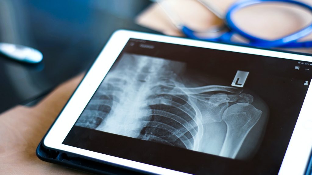
In previous blog articles, we have covered the history of radiology, inception of the DICOM standard here as well as the anatomy of the filetype and the viewer here. DICOM viewers today are packaged with a multitude of features including easy viewing and robust image post-processing. In this blog article, we will focus on understanding how these features are of merit to radiologists and how the development of computational effort has significantly impacted DICOM image viewing.
Basic Features in a DICOM Viewer
Most DICOM viewers are equipped with basic image processing features like zoom, pan, and adjusting colour, contrast and brightness. A few feature deep-dives are listed below:
Hounsfield Unit
The Hounsfield Unit (HU Scale) is used in medical imaging to quantify the radiodensity of tissues and materials. This measures the attenuation (or loss in strength) of X-rays by different parts of the body. It chooses water as the reference value of 0; high density structures (which attenuate X-rays more than water) have positive HU values, while low-density structures have negative HU values. These values help in interpreting radiology scans because regions of different HU values appear differently on the scan.
Every DICOM viewer possesses the capacity to display point-wise HU values, line-average as well as area-averaged HU values (around a rectangle/ellipse).
Draw/Annotate
DICOM viewers allow users to measure the distance between two points on the scan to allow calculation of the length of a crack/fracture or measure the size of a tumour or any other application that the radiologist wishes. Additionally, they allow viewers to calculate the area/perimeter of a specific area in the scan and measure the Cobb’s angle for spinal curvature. All these functions are a part of the ‘draw’ tool.
The DICOM Annotate tool allows radiologists to make observations about particular areas of the image, on the image itself. While other DICOM viewers provide the optional feature of including the annotated image in the report, Nandico’s DICOM viewer by-default includes annotated images in the report.
Fusion/Layout
Every DICOM viewer allows multiple layouts for medical images. They can be aligned side-by-side to a grid of images. Nandico DICOM viewer allows radiologists to view up to 9 images together in a 3×3 grid. Some DICOM viewers additionally also provide the following:
- Viewing/comparing images of same patients across different studies
- Viewing/comparing images of similar cross-sections of different patients
These are computationally expensive, and Nandico’s DICOM viewer does not support this additional functionality yet.
2DMPR
Multiplanar Reconstruction (MPR) is a tool required by radiologists to visualise images of body parts which cannot be easily examined from the base plan view. MPR allows radiologists to reconstruct images along any desired plane, providing a more comprehensive view of the anatomy.
All DICOM viewers provide easy-reconstruction along three principal planes: axial (upper/lower), sagittal (left/right) and coronal (front/back). This is called 2D-Multiplanar Reconstruction or 2D MPR.
Basic Reporting
Many DICOM viewers allow radiologists to integrate reporting with the DICOM viewer. They allow incorporating report templates that radiologists have been using, and also allow them to define shortcuts for autocompleting standardised sentences.
This is majorly available with online DICOM viewers, few offline viewers provide this functionality.
Advanced features in a DICOM Viewer
While the pre-mentioned features are available with almost every DICOM viewer, there are a few advanced features that some have developed in their portfolio to distinguish themselves.
3DMPR
While 2DMPR provides a comprehensive view along the principal axes, sometimes radiologists require to view the anatomy via oblique planes. 3D MPR tool allows them to cut the anatomy along any desired plane to provide a more detailed explanation of the spatial relationships. 3D MPR is especially valuable for visualising structures like blood vessels, airways, and joints, where a single plane may not fully reveal all details.
3DMPR is computationally expensive and online DICOM viewers seldom provide this functionality (because they risk slowing down speed considerably). However, all offline DICOM viewers (like RadiAnt, OsiriX, Weasis) provide this option.
Volume Rendering
VR in DICOM is a visualisation technique which creates a 3D image of the medical data. It utilises information from the stack of 2D images and reconstructs a 3D image from them. Different blocks of the body are highlighted in different colours allowing easy insights. Radiologists use 3D VR to identify abnormalities they might have missed (in 2D planes) as well as to plan surgeries.
3DVR is a very computationally expensive tool and no online DICOM viewer provides the same. RadiAnt’s user manual mentions that some larger CT series (>2000 images) require >1GB of active free space to be loaded successfully.
Surface Rendering
SR in DICOM is also used to create a 3D image of the medical data. However, unlike VR, SR focuses on external surfaces of organs and tissues, providing a clear view of their shapes and contours. The model is represented as a mesh of interconnected triangles, forming a surface that approximates the external shape of the structures. Radiologists use SR to guide catheter placements with precision, design customised prosthetics and plan surgeries.
Surface Rendering is also a very computationally expensive tool and no online DICOM viewer provides the same.
MIP & MinIP
MIP (maximum intensity projection) displays the maximum pixel intensity (brightest pixel) along a specific projection ray or line in a 3D dataset. MIP pictures are useful for highlighting vascular structures.
MinIP (minimum intensity projection)displays the minimum pixel intensity (darkest pixel) along each projection ray in a 3D dataset. MinIP pictures are useful for highlighting air-filled structures like bronchial tree in lungs.
HL7 Integration
HL7 (Health-level 7) is an international standard used for interfacing DICOM viewers and the whole PACS system to the hospital information system (HIS) or the radiology information system (RIS). Not all DICOM viewers support this.
Report Dictation
One prominent feature, which paves the way for AI in radiology is the concept of speech-to-text or report dictation. DICOM viewers which have reporting features integrated sometimes allow radiologists to dictate their reports. This requires highly trained models which can recognise various accents as well as produce medically-correct terminology. Augnito is one such tool which allows report dictation. Not all viewers have this capability and is thus classified under advanced features.
How has computational efficiency paved the way for robust viewers?
DICOM viewers have been greatly benefited due to improvements in computational efficiency. A few features that have been enabled only because of disruptions are:
- 3DVR and 3DMPR, which were not possible earlier
- Image fusion, which involves comparing images across modalities
- AI models for voice detection, computer vision for image enhancement and anomaly detection
- The concept of cloud-based PACS and web-based DICOM viewers was possible only because of computational efficiency
How does Nandico’s DICOM Viewer fin-in?
Nandico’s DICOM viewer is a cloud-based viewer enabling remote-access and user-friendly mobile-first solutions. It possesses all basic DICOM viewer functionality as mentioned above and a couple advanced features are in the pipeline (stay tuned!) Additionally, we offer a clean and intuitive UI, and a transparent pricing plan. Head over to Contact – Nandico to get a free demo now!
The Bottomline
DICOM viewers across the world have various features that enable radiologists to diagnose various ailments in a simple, intuitive, yet powerful format. More recently there has been a tradeoff- providing cloud-based PACS to prioritise mobility and remote access, but giving up on computationally expensive features like 3D MPR, VR and SR; or vice versa. We promise to continue this discussion in the upcoming articles. Till then, stay tuned!
